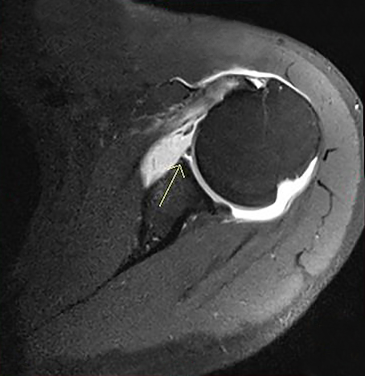
AIM Offers MRI Arthrograms
AIM Medical Imaging offers MRI arthrograms of the hip and shoulders–but what do these procedures entail? Arthrograms are used especially for imaging joints, and are often indicated after a patient suffers a traumatic injury such as an auto accident. While an arthrogram may also be performed through X-Ray or CT, MRI is ideal because it will not expose the patient to ionizing radiation.
What is the Arthrogram Process?
First, the patient must sign a screening form stating that they are not taking blood thinners, allergic to local anesthetics, or have any other health issues that may put them at risk when receiving an arthrogram.
In order to see if there is an injury or tear in a joint’s cartilage, gadolinium contrast must first be injected directly into the joint. At AIM, our radiologists use ultrasound imaging to pinpoint the exact location for the injection site. Other clinics may use low resolution X-Ray in order to find the joint, but AIM guarantees that your arthrogram is 100 per cent free of ionizing radiation. Once the joint is located, AIM’s radiologist freezes the area with a local anesthetic before injecting the contrast into the joint with a long, sterilized needle. Once contrast has been injected, the patient is instructed to gently move the area–be it the shoulder or hip–in order to allow the contrast to travel throughout the joint.
How Does it Work?
If the joint is indeed torn or damaged, contrast will reveal where. The contrasted (white area) in the above AIM arthrogram image shows an anterioinferior labral tear with associated soft tissue Bankart and bony Hill-Sachs deformity. Put simply, a rupture in the joint is clearly illustrated with the white contrast materials bleeding out of it.
AIM arthrograms are done on-site, overseen by qualified radiologists and technologists. Like all MRI scans done at AIM Medical Imaging, patients can expect to receive their results in a face-to-face consultation with our radiologist immediately following the procedure.

