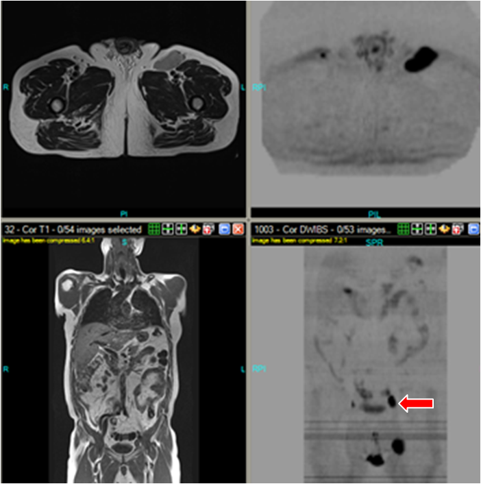
Whole Body Diffusion MRI in the Journal of Magnetic Resonance Imaging – Part One
Last week we announced the publication of Dr. Attariwala’s and Wayne’s article in the Journal of MRI. This week, let’s take a look at their discoveries, and how they may be applied to diagnostic medicine to advance the state of the art for patients’ best interests.
In the article, authors Attariwala and Picker share their observations gleaned from scanning hundreds of patients at AIM Medical Imaging with whole body DWIBS techniques, as well as theory, equations, and tips for optimized image viewing.
 Diffusion Weighted Techniques are so powerful because they enable imaging of movement and/or blockage of water at the cellular level. This explains why DWI is integral for tumour staging and lesion detection; even in the earliest stages of tumour formation, DWI techniques will pick up a resistance of water movement–often early enough to put a halt to the onset of cancer! Of DWI, Dr. Attariwala states, “This functional technique has long been used in the brain to demonstrate alteration in intra- and extra cellular water content from disruption of the transmembrane water flux. These cellular level alterations are visible before changes can be identified on morphologic routine sequences.” So, while DWI is not a novel technique, Whole Body Diffusion MRI is, and Attariwala and Picker have outlined the reasons which sets AIM’s technique apart from traditional CT/PET scans; not least of these is that MRI is radiation free.
What else can Whole Body Diffusion MRI do? Attariwala and Picker’s article states it can:
-observe multiple myeloma (bone marrow cancer), lymphoma (blood cancer) and skeletal metastases
-detect hemorrhagic degradation products, infections/abcesses, colitis inflammation
-easily distinguish between tumours, lesions, free fluids and empyema (pus)
-gauge to what extent a diseased patient has responded to treatments/therapies/surgeries/medications
-identify other abnormalities outside of lesions or tumours
Stay tuned for more exclusive posts in the coming weeks, including a Q & A with Dr. Attariwala!

