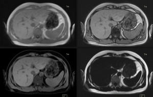
New Research finds MRI Superior to CT for Liver Cancer Diagnosis
A new study published in the Radiological Society of North America (RSNA) journal Radiology has reviewed and analyzed the contrasting abilities of MRI and CT scanning of the liver for cancer.
 The verdict is in: MRI is superior to CT for cancer diagnosis of the liver. And a victory for MRI is, as always, a positive step for patients everywhere because it means less overall exposure to ionizing radiation–an inherent risk of CT imaging.
The Korean study, entitled Hepatocellular Carcinoma (HCC): Diagnostic Performance of Multidetector CT and MR Imaging–a Systematic Review and Meta Analysis compared 3,924 patient case studies of chronic liver disease. The researchers found MRI showed superior lesion sensitivity when imaging the liver, enabling them to more accurately diagnose HCC (the most common type of liver cancer) characteristics within pre-existing liver disease conditions. Overall, the lesion-detection sensitivity for MRI ranked at 79 per cent, while CT’s sensitivity sat at 72 per cent. Contrast-aided MRI scans attained an even higher sensitivity ranking. Notably, the research was limited to patients with small tumours.
As the researchers concluded: “MR imaging showed higher per-lesion sensitivity than multidetector CT and should be the preferred imaging modality for the diagnosis of HCCs in patients with chronic liver disease.”
Related reading from the AIM blog:
When Should You Choose MRI over CT, PET or Ultrasound?
The Importance of an Onsite MRI Radiologist for Diagnostic Accuracy

